
| Synonyms | |
| Status | |
| Molecule Category | Free-form |
| UNII | LDR3USH1NJ |
| EPA CompTox | DTXSID90196103 |
Structure
| InChI Key | HQSBCDPYXDGTCL-UHFFFAOYSA-N |
|---|---|
| Smiles | |
| InChI |
|
Physicochemical Descriptors
| Property Name | Value |
|---|---|
| Molecular Formula | C16H15N7O |
| Molecular Weight | 321.34 |
| AlogP | 2.0 |
| Hydrogen Bond Acceptor | 8.0 |
| Hydrogen Bond Donor | 2.0 |
| Number of Rotational Bond | 3.0 |
| Polar Surface Area | 121.67 |
| Molecular species | NEUTRAL |
| Aromatic Rings | 4.0 |
| Heavy Atoms | 24.0 |
Pharmacology
| Targets | EC50(nM) | IC50(nM) | Kd(nM) | Ki(nM) | Inhibition(%) | |
|---|---|---|---|---|---|---|
|
Membrane receptor
Family A G protein-coupled receptor
Small molecule receptor (family A GPCR)
Nucleotide-like receptor (family A GPCR)
Adenosine receptor
|
- | - | 0.631-100 | 1.3-68 | - |
 Homo sapiens
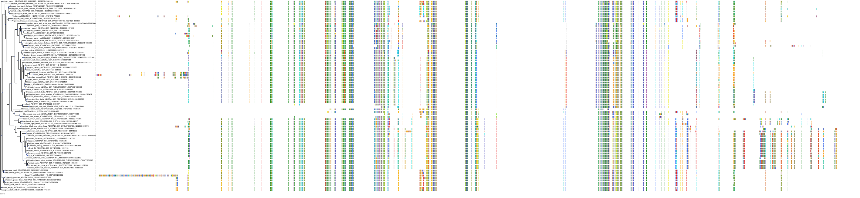
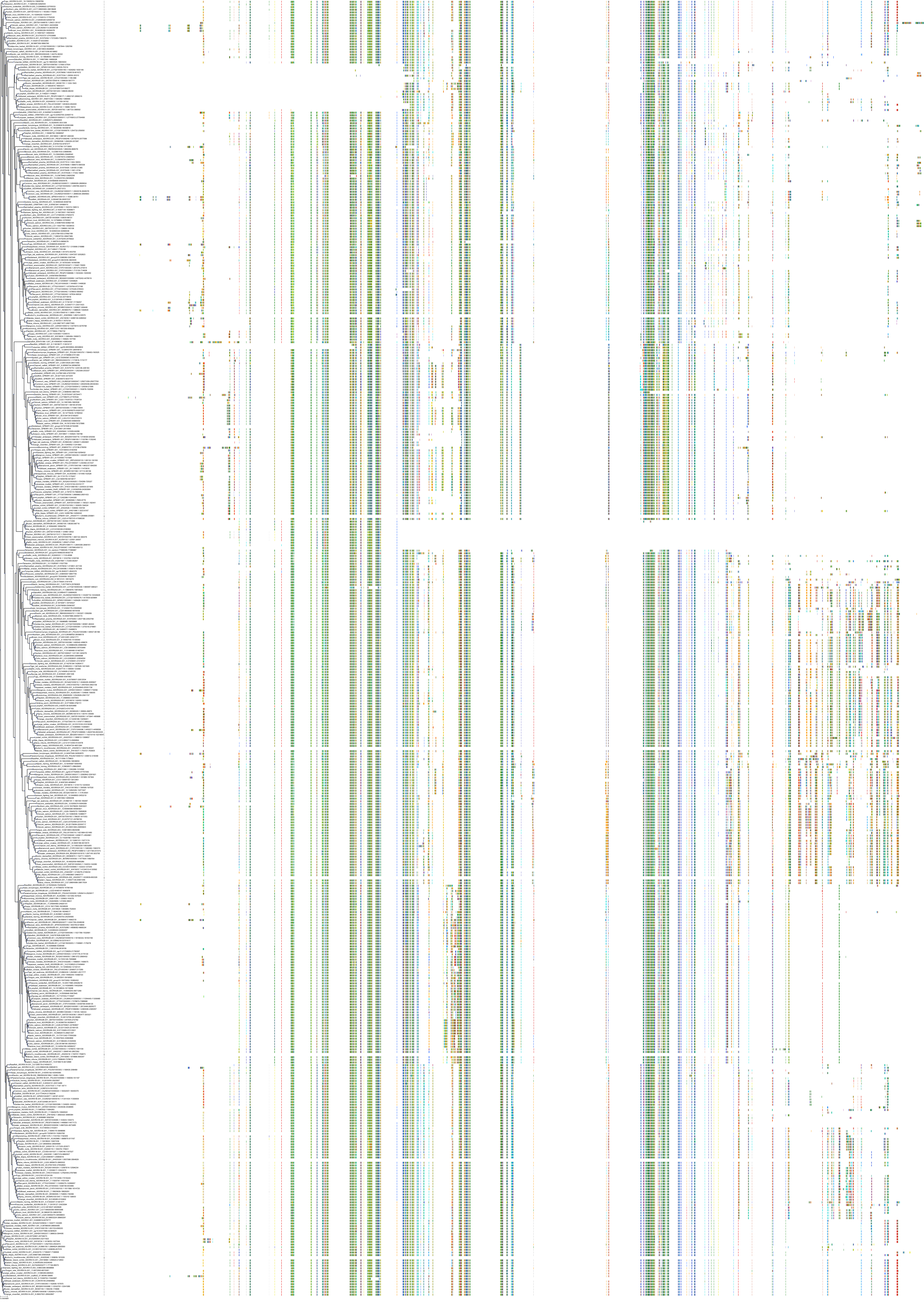
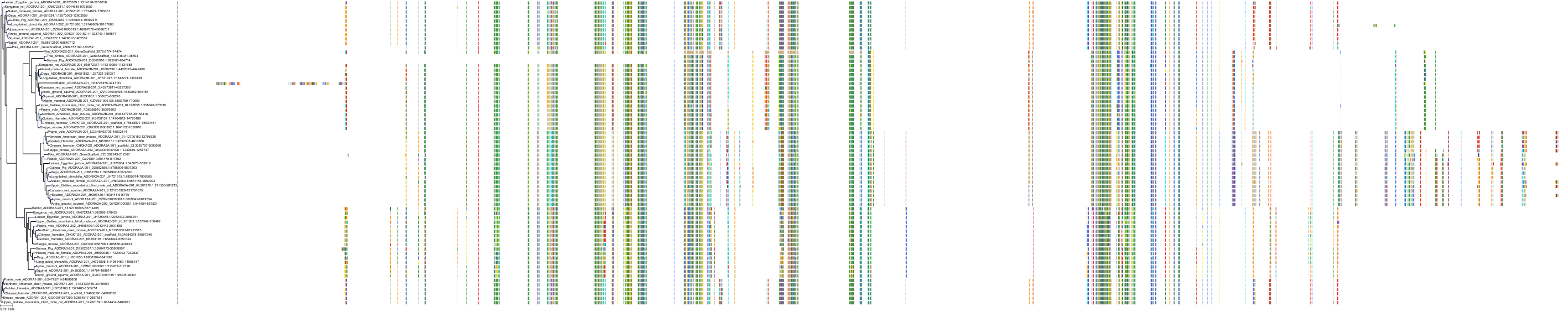
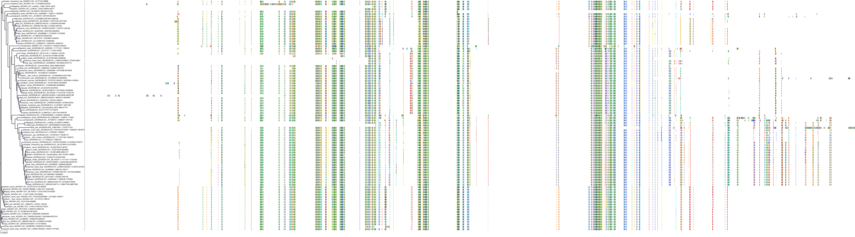
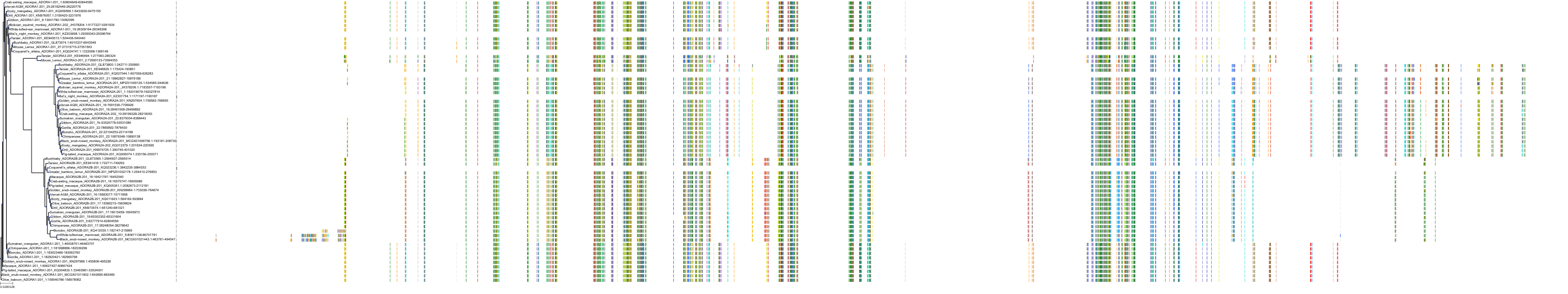
Target Conservation
|
Protein: Adenosine A2a receptor Description: Adenosine receptor A2a Organism : Homo sapiens P29274 ENSG00000128271 |
|
|||


Related Entries

Cross References
| Resources | Reference |
|---|---|
| CAS NUMBER | 442908-10-3 |
| ChEMBL | CHEMBL447664 |
| DrugBank | DB06625 |
| FDA SRS | LDR3USH1NJ |
| Guide to Pharmacology | 5612 |
| PDB | 9XT |
| PubChem | 21874557 |
| SureChEMBL | SCHEMBL2849027 |
| ZINC | ZINC000040863182 |
CONTENTS
