
Structure
| InChI Key | MWKYMZXCGYXLPL-ZDUSSCGKSA-N |
|---|---|
| Smiles | |
| InChI |
|
Physicochemical Descriptors
| Property Name | Value |
|---|---|
| Molecular Formula | C21H25F3N6O2 |
| Molecular Weight | 450.47 |
| AlogP | 2.88 |
| Hydrogen Bond Acceptor | 7.0 |
| Hydrogen Bond Donor | 1.0 |
| Number of Rotational Bond | 5.0 |
| Polar Surface Area | 83.48 |
| Molecular species | NEUTRAL |
| Aromatic Rings | 2.0 |
| Heavy Atoms | 32.0 |
Pharmacology
| Mechanism of Action | Action | Reference |
|---|---|---|
| PI3-kinase p110-delta subunit inhibitor | INHIBITOR | PubMed PubMed |
| Targets | EC50(nM) | IC50(nM) | Kd(nM) | Ki(nM) | Inhibition(%) | |
|---|---|---|---|---|---|---|
|
Enzyme
Kinase
Protein Kinase
AGC protein kinase group
AGC protein kinase RSK family
AGC protein kinase MSK subfamily
|
- | - | - | - | 76 | |
|
Enzyme
Kinase
Protein Kinase
Atypical protein kinase group
Atypical protein kinase PIKK family
|
- | 880 | - | - | - | |
|
Enzyme
Kinase
Protein Kinase
CAMK protein kinase group
CAMK protein kinase RSKb family
CAMK protein kinase MSKb subfamily
|
- | - | - | - | 76 | |
|
Enzyme
Transferase
|
- | 7-424 | - | - | - |
 Homo sapiens
 Macaca fascicularis
 Mus musculus
 Rattus norvegicus
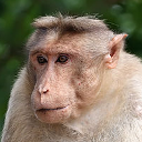
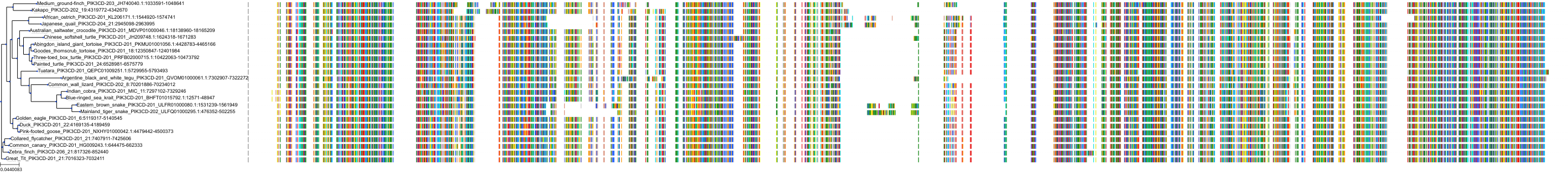
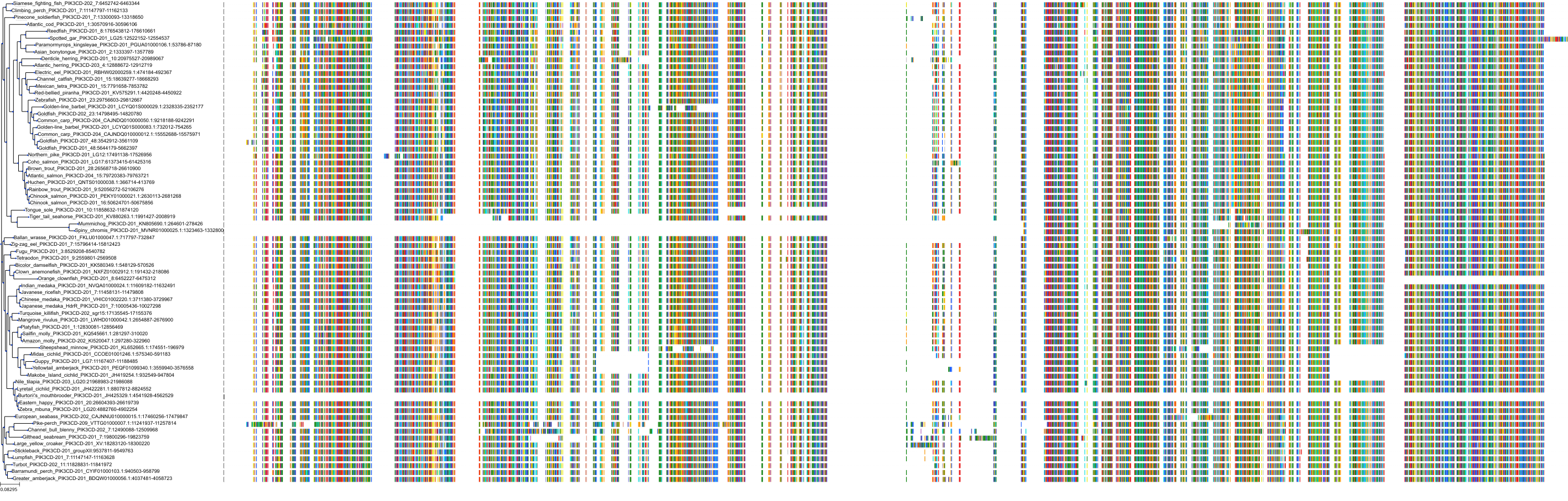
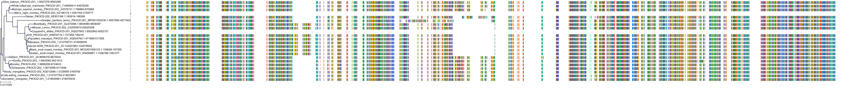
Target Conservation
|
Protein: PI3-kinase p110-delta subunit Description: Phosphatidylinositol 4,5-bisphosphate 3-kinase catalytic subunit delta isoform Organism : Homo sapiens O00329 ENSG00000171608 |
|
|||


Cross References
| Resources | Reference |
|---|---|
| CAS NUMBER | 1354690-24-6 |
| ChEMBL | CHEMBL3643413 |
| DrugBank | DB16217 |
| FDA SRS | L22772Z9CP |
| Guide to Pharmacology | 9424 |
| PDB | 9NQ |
| PubChem | 57495353 |
| SureChEMBL | SCHEMBL323054 |
CONTENTS
