
| Synonyms | |
| Status | |
| Molecule Category | Free-form |
| UNII | J1I0P6WT7T |
| EPA CompTox | DTXSID301020622 |
Structure
| InChI Key | YHYKUSGACIYRML-KRWDZBQOSA-N |
|---|---|
| Smiles | |
| InChI |
|
Physicochemical Descriptors
| Property Name | Value |
|---|---|
| Molecular Formula | C17H17F2N5O3S |
| Molecular Weight | 409.42 |
| AlogP | 1.63 |
| Hydrogen Bond Acceptor | 5.0 |
| Hydrogen Bond Donor | 3.0 |
| Number of Rotational Bond | 3.0 |
| Polar Surface Area | 115.25 |
| Molecular species | NEUTRAL |
| Aromatic Rings | 2.0 |
| Heavy Atoms | 28.0 |
Pharmacology
| Targets | EC50(nM) | IC50(nM) | Kd(nM) | Ki(nM) | Inhibition(%) | |
|---|---|---|---|---|---|---|
|
Enzyme
Protease
Aspartic protease
Aspartic protease AA clan
Aspartic protease A1A subfamily
|
- | 0.38-85 | - | 0.4-10.9 | 95.04-98.6 |
 Canis lupus familiaris
 Homo sapiens
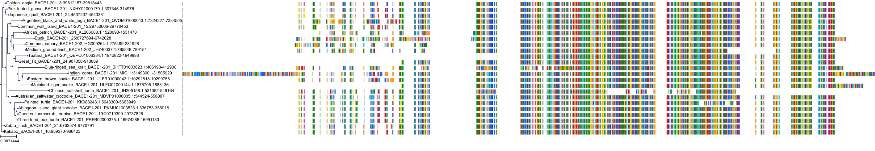
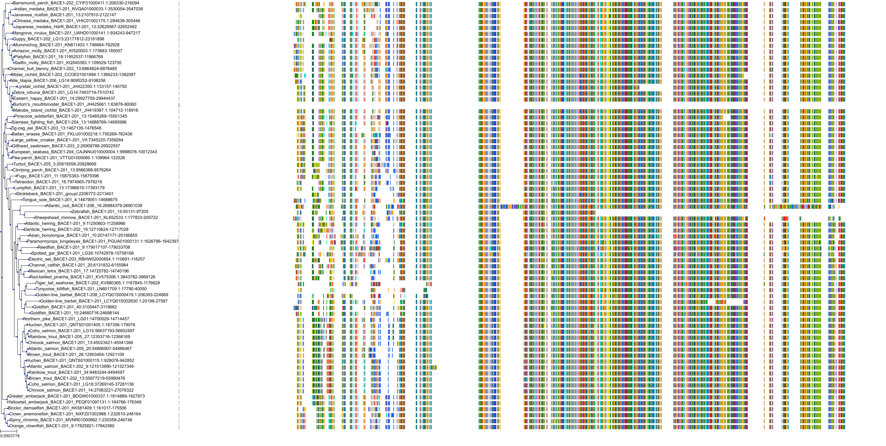
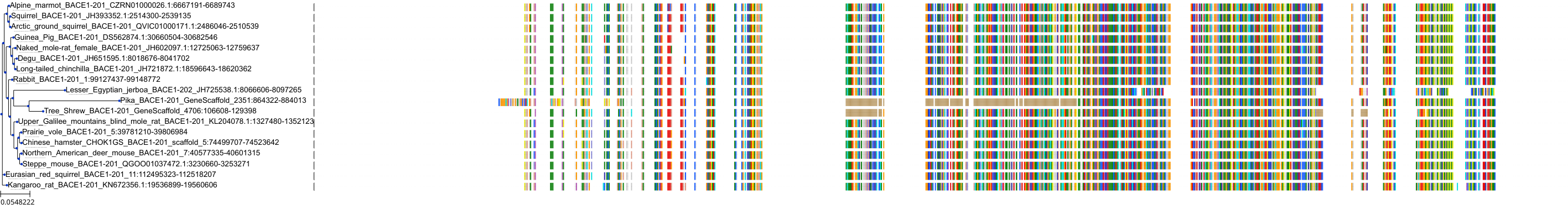
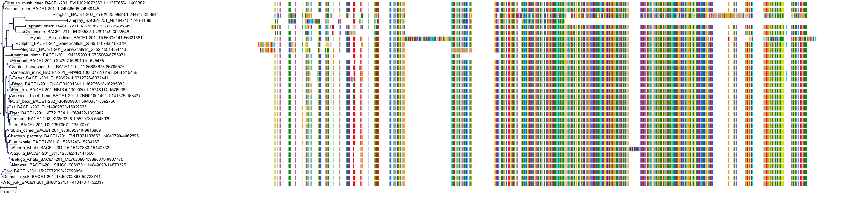
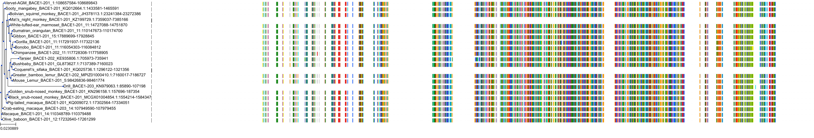
Target Conservation
|
Protein: Beta-secretase 1 Description: Beta-secretase 1 Organism : Homo sapiens P56817 ENSG00000186318 |
|
|||


Cross References
| Resources | Reference |
|---|---|
| CAS NUMBER | 1286770-55-5 |
| ChEMBL | CHEMBL3301601 |
| DrugBank | DB12285 |
| FDA SRS | J1I0P6WT7T |
| Guide to Pharmacology | 8699 |
| PDB | 66F |
| PubChem | 51352361 |
| SureChEMBL | SCHEMBL10328722 |
| ZINC | ZINC000144542146 |
CONTENTS
