
Structure
| InChI Key | GTUIQNHJSXQMKW-UHFFFAOYSA-N |
|---|---|
| Smiles | |
| InChI |
|
Physicochemical Descriptors
| Property Name | Value |
|---|---|
| Molecular Formula | C11H13ClN2O2 |
| Molecular Weight | 240.69 |
| AlogP | 1.22 |
| Hydrogen Bond Acceptor | 3.0 |
| Hydrogen Bond Donor | 1.0 |
| Number of Rotational Bond | 1.0 |
| Polar Surface Area | 45.48 |
| Molecular species | BASE |
| Aromatic Rings | 1.0 |
| Heavy Atoms | 16.0 |
Pharmacology
| Mechanism of Action | Action | Reference |
|---|---|---|
| Neuronal acetylcholine receptor; alpha4/beta2 agonist | AGONIST | PubMed |
| Targets | EC50(nM) | IC50(nM) | Kd(nM) | Ki(nM) | Inhibition(%) | |
|---|---|---|---|---|---|---|
|
Ion channel
Ligand-gated ion channel
5HT3 receptor
|
- | - | - | 700 | 87-97 | |
|
Ion channel
Ligand-gated ion channel
Nicotinic acetylcholine receptor
Nicotinic acetylcholine receptor alpha subunit
|
- | - | - | 30-34 | - | |
|
Ion channel
Ligand-gated ion channel
Nicotinic acetylcholine receptor
Nicotinic acetylcholine receptor beta subunit
|
- | - | - | 30-34 | - |
 Homo sapiens
 Mus musculus
 Rattus norvegicus
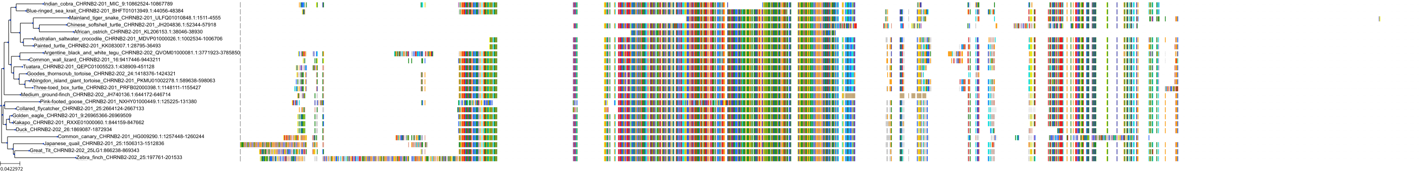
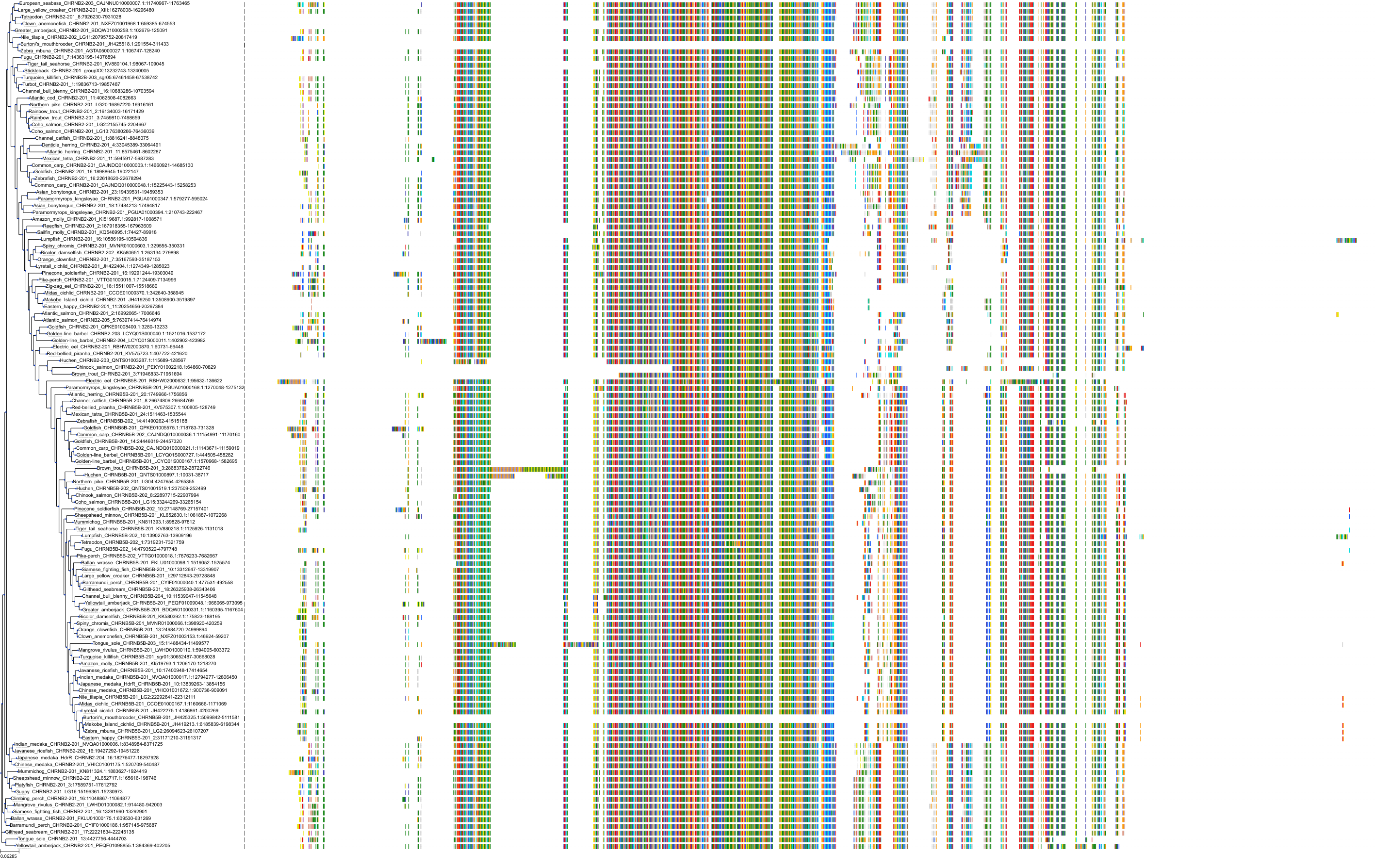
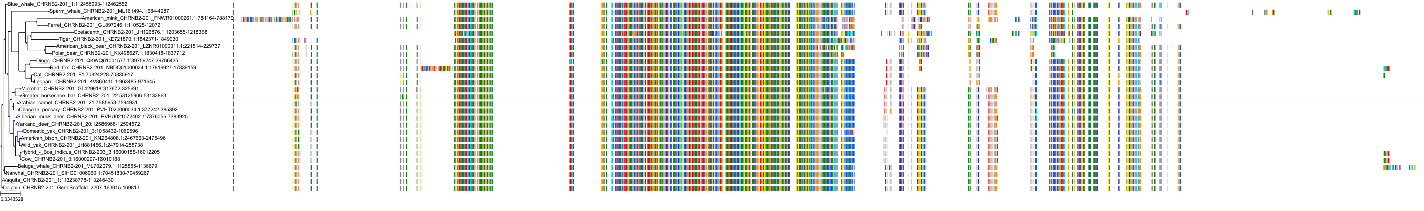
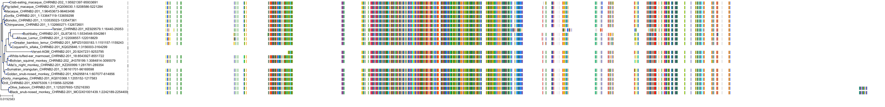
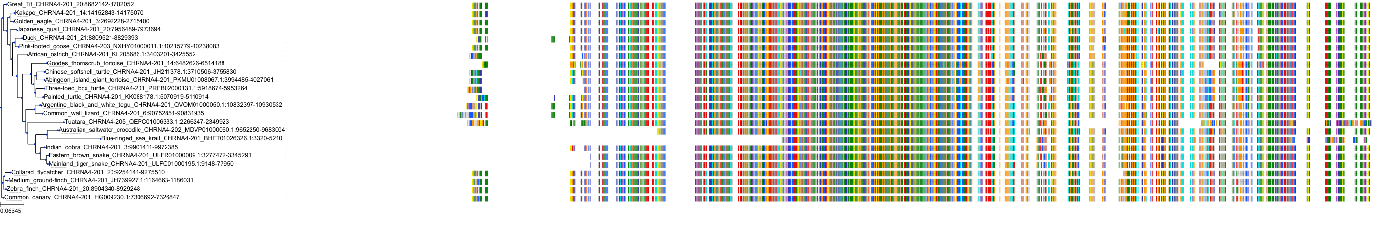
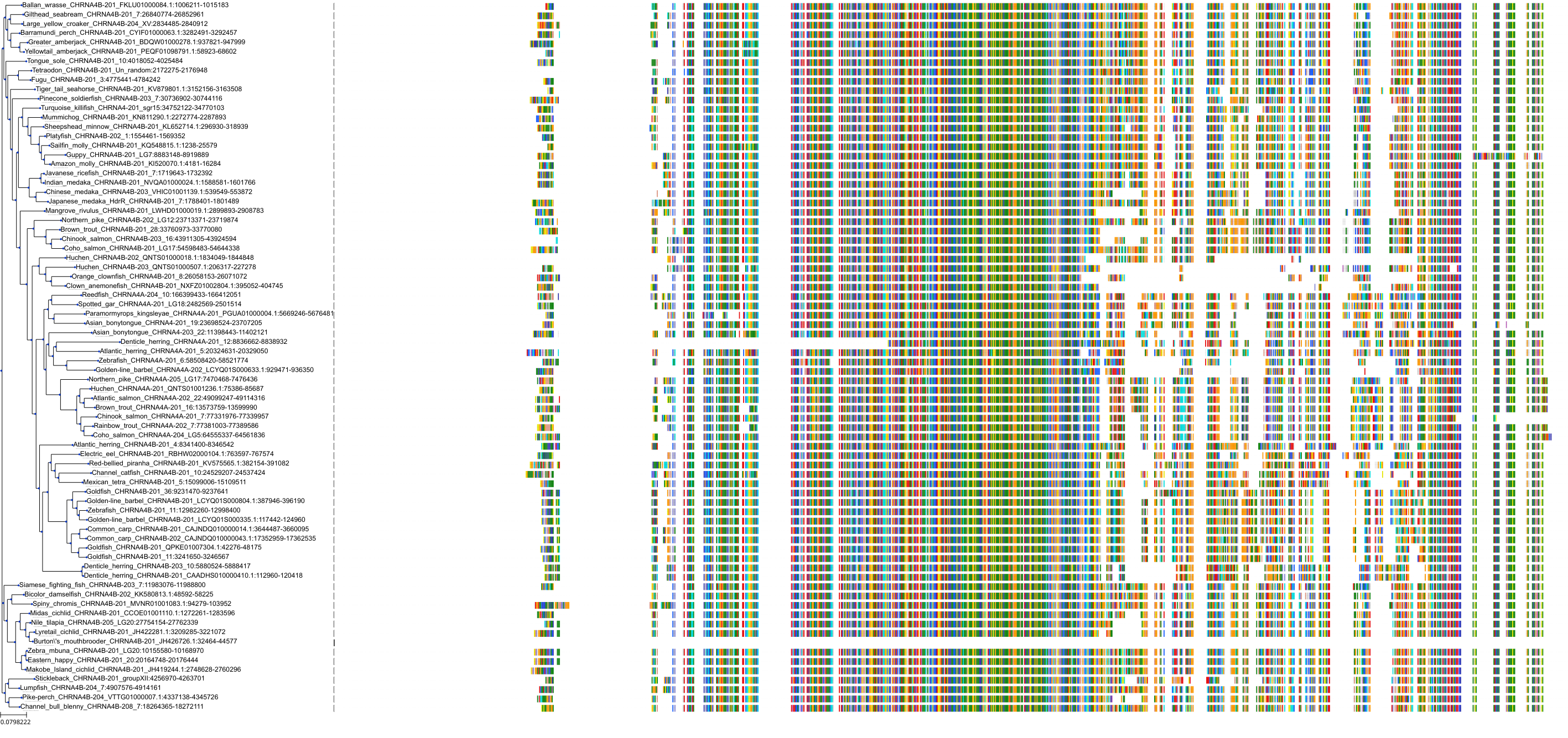
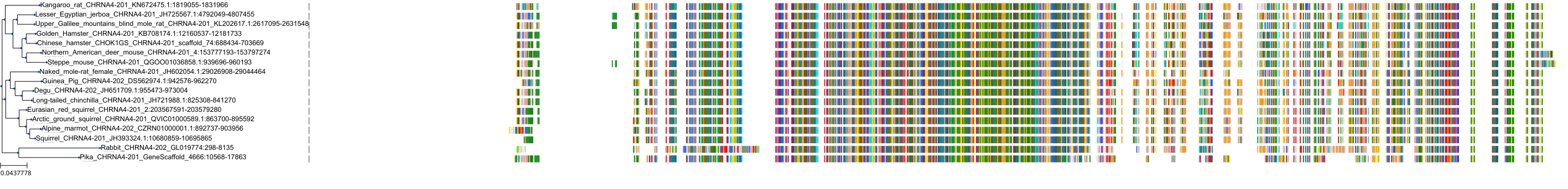
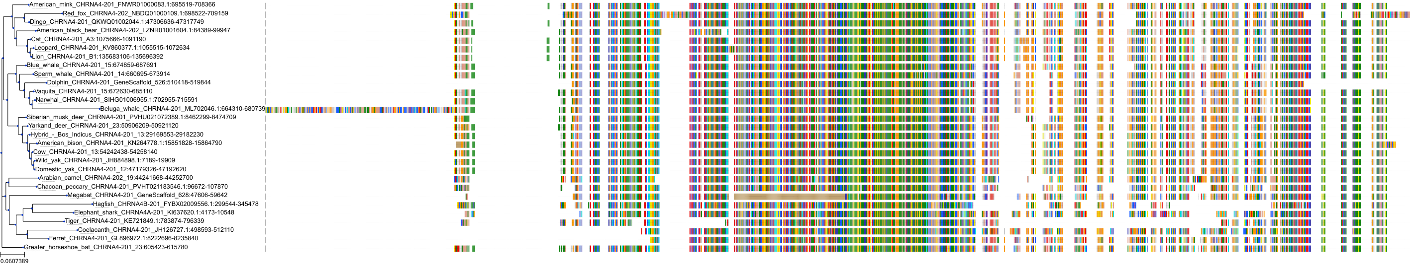
Target Conservation
|
Protein: Neuronal acetylcholine receptor; alpha4/beta2 Description: Neuronal acetylcholine receptor subunit beta-2 Organism : Homo sapiens P17787 ENSG00000160716 |
|
|||
|
Protein: Neuronal acetylcholine receptor; alpha4/beta2 Description: Neuronal acetylcholine receptor subunit alpha-4 Organism : Homo sapiens P43681 ENSG00000101204 |
|
|||


Cross References
| Resources | Reference |
|---|---|
| CAS NUMBER | 1788894-06-3 |
| ChEMBL | CHEMBL2179529 |
| FDA SRS | ANR9OP1V17 |
| SureChEMBL | SCHEMBL584395 |
CONTENTS
