
Structure
| InChI Key | HNLRRJSKGXOYNO-UHFFFAOYSA-N |
|---|---|
| Smiles | |
| InChI |
|
Physicochemical Descriptors
| Property Name | Value |
|---|---|
| Molecular Formula | C23H26N6O3S |
| Molecular Weight | 466.57 |
| AlogP | 2.59 |
| Hydrogen Bond Acceptor | 9.0 |
| Hydrogen Bond Donor | 2.0 |
| Number of Rotational Bond | 6.0 |
| Polar Surface Area | 107.01 |
| Molecular species | NEUTRAL |
| Aromatic Rings | 4.0 |
| Heavy Atoms | 33.0 |
Pharmacology
| Mechanism of Action | Action | Reference |
|---|---|---|
| Fibroblast growth factor receptor inhibitor | INHIBITOR | DOI |
Target Conservation
|
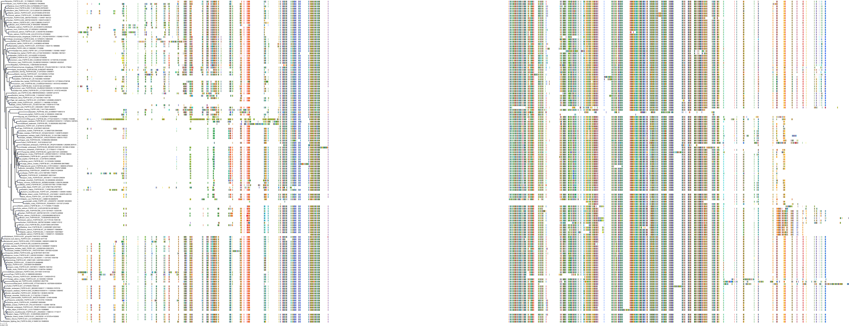
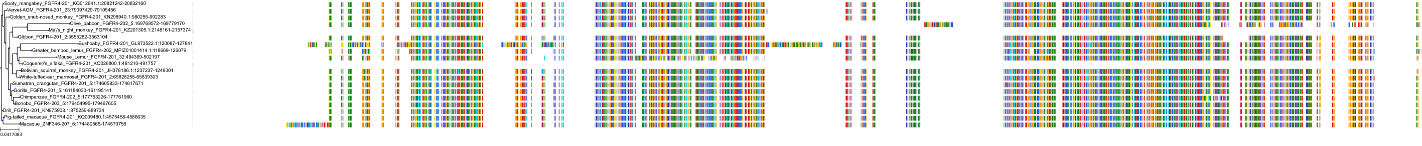
Protein: Fibroblast growth factor receptor Description: Fibroblast growth factor receptor 1 Organism : Homo sapiens P11362 ENSG00000077782 |
|
|||
|
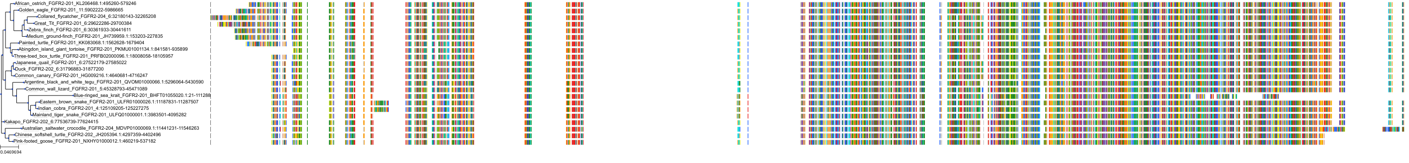
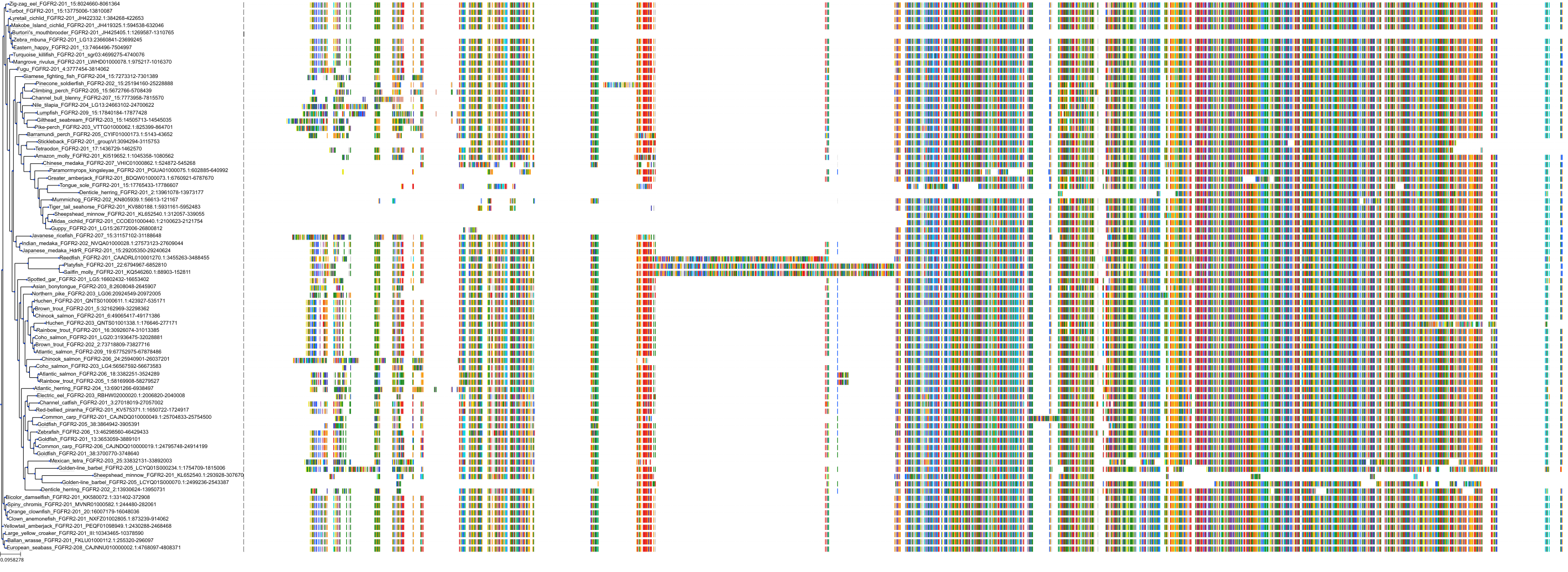
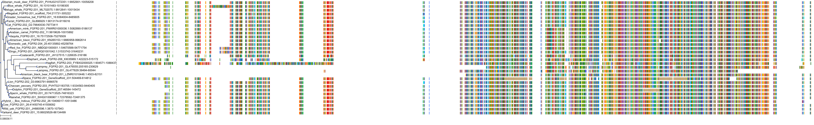
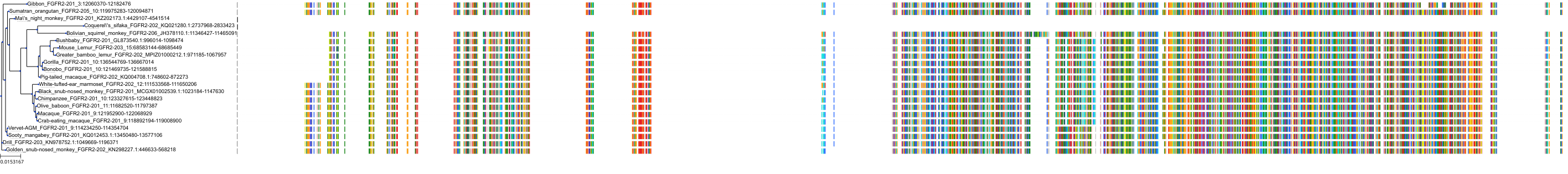
Protein: Fibroblast growth factor receptor Description: Fibroblast growth factor receptor 2 Organism : Homo sapiens P21802 ENSG00000066468 |
|
|||
|
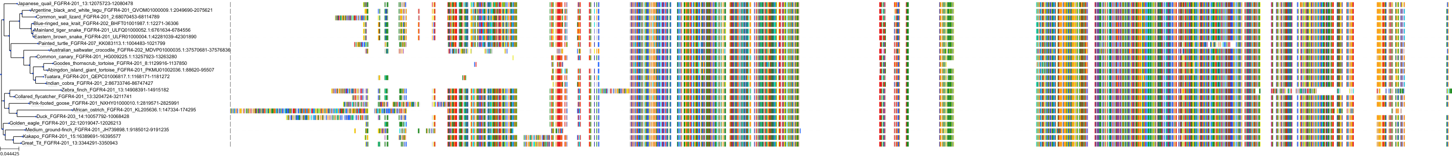
Protein: Fibroblast growth factor receptor Description: Fibroblast growth factor receptor 4 Organism : Homo sapiens P22455 ENSG00000160867 |
|
|||
|
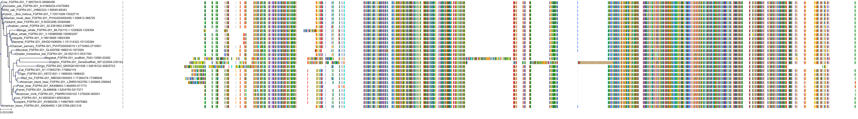
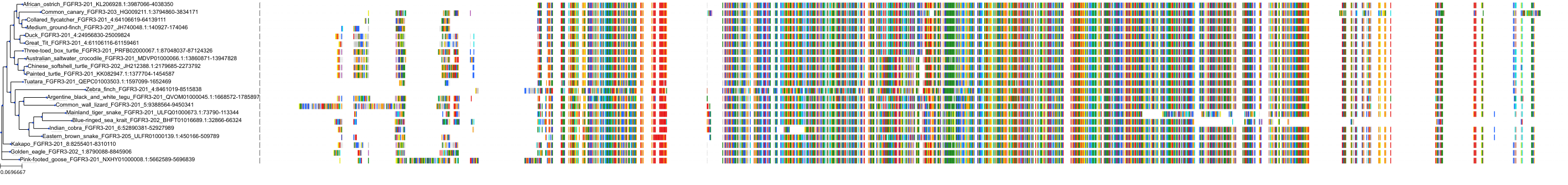
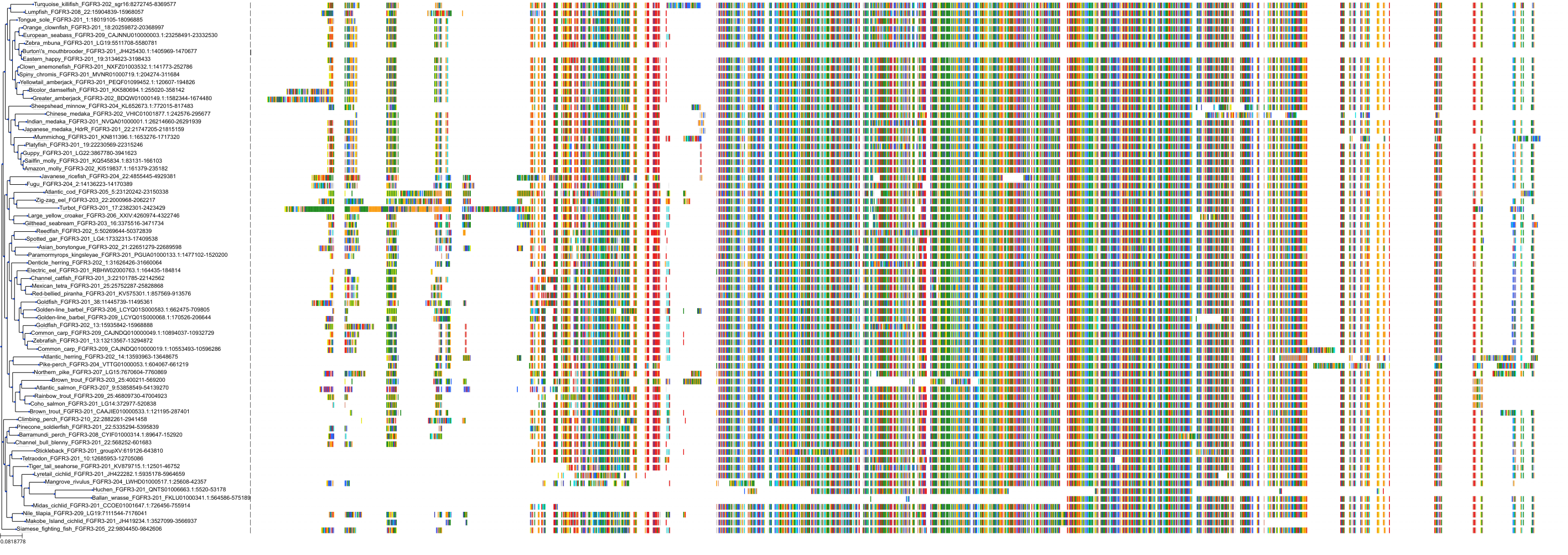
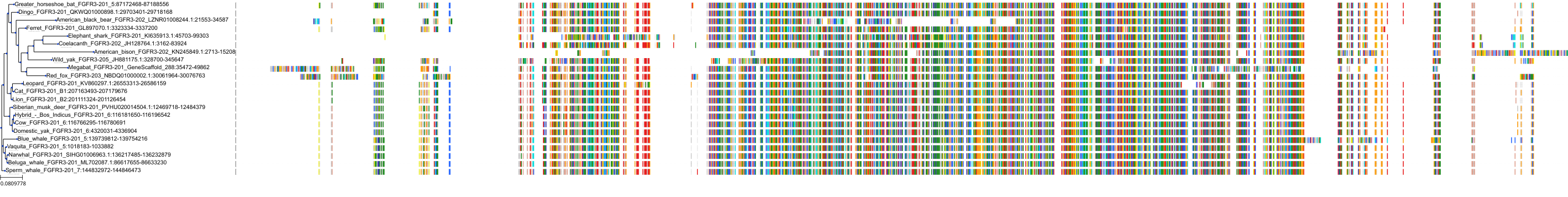
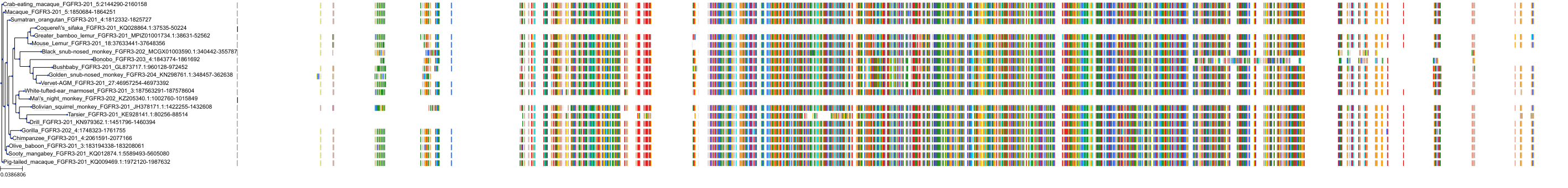
Protein: Fibroblast growth factor receptor Description: Fibroblast growth factor receptor 3 Organism : Homo sapiens P22607 ENSG00000068078 |
|
|||


Cross References
| Resources | Reference |
|---|---|
| CAS NUMBER | 1443530-05-9 |
| ChEMBL | CHEMBL3963485 |
| DrugBank | DB15078 |
| FDA SRS | 98BSN6N516 |
| Guide to Pharmacology | 9789 |
| PubChem | 71611869 |
| SureChEMBL | SCHEMBL15023004 |
CONTENTS
