
Structure
| InChI Key | CWFVVQFVGMFTBD-SECBINFHSA-N |
|---|---|
| Smiles | |
| InChI |
|
Physicochemical Descriptors
| Property Name | Value |
|---|---|
| Molecular Formula | C18H13ClF4N6O |
| Molecular Weight | 440.79 |
| AlogP | 3.63 |
| Hydrogen Bond Acceptor | 6.0 |
| Number of Rotational Bond | 2.0 |
| Polar Surface Area | 76.8 |
| Molecular species | NEUTRAL |
| Aromatic Rings | 3.0 |
| Heavy Atoms | 30.0 |
Pharmacology
| Mechanism of Action | Action | Reference |
|---|---|---|
| P2X purinoceptor 7 antagonist | ANTAGONIST | ClinicalTrials PubMed PubMed PubMed |
| Targets | EC50(nM) | IC50(nM) | Kd(nM) | Ki(nM) | Inhibition(%) | |
|---|---|---|---|---|---|---|
|
Ion channel
Ligand-gated ion channel
P2X receptor
|
- | 1.549-31.62 | - | 5.012-5.012 | - |
 Canis lupus familiaris
 Homo sapiens
 Macaca mulatta
 Mus musculus
 Rattus norvegicus
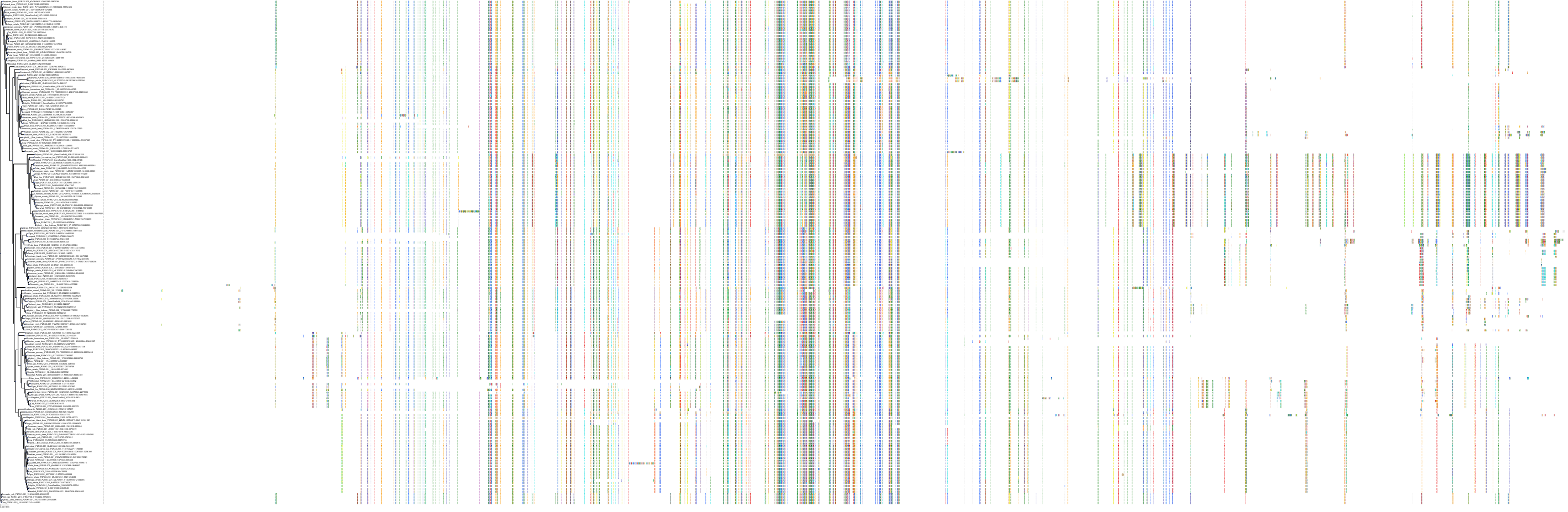
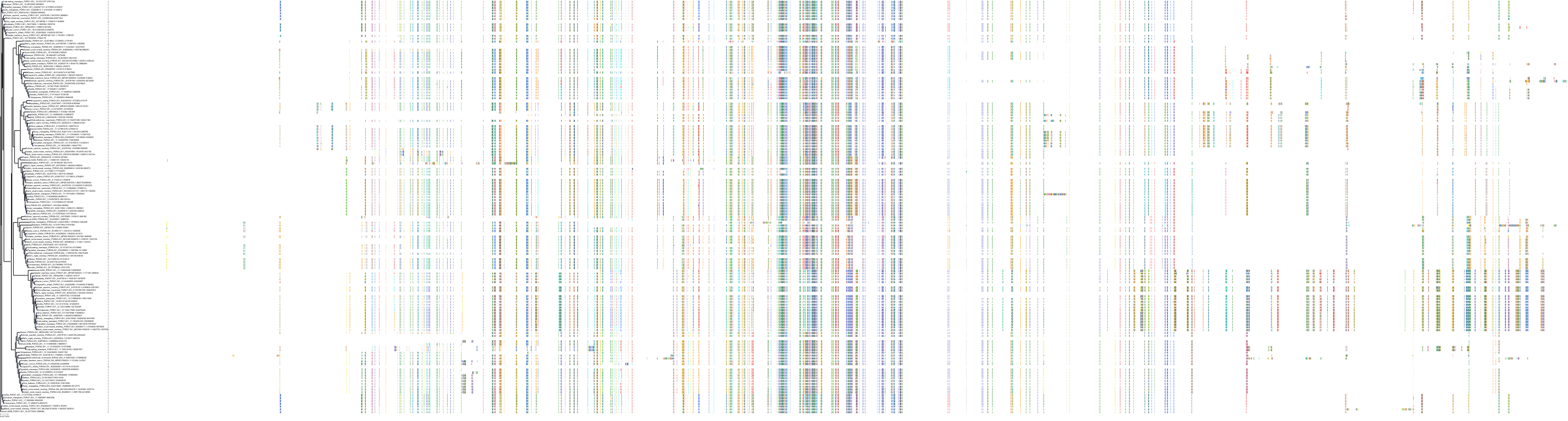
Target Conservation
|
Protein: P2X purinoceptor 7 Description: P2X purinoceptor 7 Organism : Homo sapiens Q99572 ENSG00000089041 |
|
|||


Cross References
| Resources | Reference |
|---|---|
| CAS NUMBER | 1627902-21-9 |
| ChEMBL | CHEMBL4079239 |
| DrugBank | DB15358 |
| FDA SRS | 32524GLF40 |
| PubChem | 90409366 |
| SureChEMBL | SCHEMBL16036477 |
CONTENTS
